Sunday, September 24, 2017

Brugada syndrome - types

Look at the image, then follow the text.

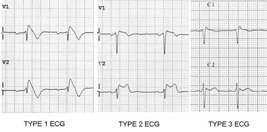
Look at the right pericordial leads (V1-V3)
Brugada type 1: 
1) Coved type ST elevation
2) J point or ST elevation > 2mm
3) Negative T wave
Brugada type 2: 
1) saddle-back appearance
2) J point elevation > 2mm, ST elevation > 1mm
3) Positive or Bipshasic T wave
Brugada type 3:
1) saddleback or coved appearance
2) ST-segment elevation <1 mm


Tuesday, March 28, 2017

Tilt-table test

  1. is used to indentify cause of SENCOPE
  2. we will identify cause of sencope: vasodepressor or cardioinhibitory
  3. patient lay on table in the supin position
  4. tilting the table maximum: 60-80degrees for 25-40minute
  5. What is result? - Nothing? Lets repeat test with drugs which provoke sencope.
  6. Which drugs we can use? - sublingual nitroglycerin, epinephrine, adenosine, mostly-isoproterenol
  7. Role of isoproterenol: - induces a vasodepressor (decrease in heart rate and blood pressure)
  8. Neurocardyogenic sencopes: Vasodepressor, Carboinhibitory, Mixed
  9. What results can be?: Autonomic failure, Postural Tachycardia, Vasodepresor,
    RESPONSES

    a normal response is an early, slight drop in BP with a compensatory increase in HR mediated by the autonomic nervous system.


    With autonomic dysfunction, a progressive fall in BP is not counteracted by an increase in HR

    In postural tachycardia syndrome, an exaggerated increase in HR is seen


    Vasodepressor is a type of neurocardiogenic sencope. Pure vasodepressor response is a relatively sudden drop in BP without a marked change in HR

    cardioinhibitory is also neurocadiogenic sencope. Cardioinhibitory response shows a sudden decrease in HR without a change in BP

    A mixed response shows decreases in both HR and BP.(also neurocardiogenic sencope)

Sunday, February 26, 2017

Why we review the serum creatinine?

Serum creatinine

1) Liver produce creatinine
2) Creatin kinase ferment phosphorylate creatinine to: phosphocreatine
3) phosphocreatine is high-energy compound so muscle and brain need it mainly
4) at the end of metabolysm kidnet remove creatinin
5) if the function of kidneys funstion slows down then creatinine level will increase

Do not be envious! Share your knowledge with us. Comment more information. Share also..

Right parasternal position

Do you use right parasternal position on echocardiography? If we dont use then why we say that position while talking about echocardiography?

This position is useful to examine the aorta or interatrial septum and is also useful in patients with congenital malposition of the heart, such as dextrocardia. If you have more information do not be envious. Share your knowledge with us.

Friday, February 24, 2017

Congenital Heart Defects

Lets put the list of CHD here, then step by step review them :)
Aortic stenosis
Atrial septal defect (ASD)
Atrioventricular septal defect (AVSD)
Bicuspid aortic valve
Dextrocardia
Double inlet left ventricle (DILV)
Double outlet right ventricle (DORV)
Ebstein's anomaly
Hypoplastic left heart syndrome (HLHS)
Hypoplastic right heart syndrome (HRHS)
Mitral stenosis
Pulmonary atresia
Pulmonary stenosis
Transposition of the great vessels
dextro-Transposition of the great arteries (d-TGA)
levo-Transposition of the great arteries (l-TGA)
Tricuspid atresia
Persistent truncus arteriosus
Ventricular septal defect (VSD)
Wolff-Parkinson-White syndrome (WPW)
Some conditions affect the great vessels or other vessels in close proximity to the heart, but not the heart itself, but are often classified as congenital heart defects.
Coarctation of the aorta (CoA)
Double aortic arch, aberrant subclavian artery, and other malformations of the great arteries
Interrupted aortic arch (IAA)
Patent ductus arteriosus (PDA)
Scimitar syndrome (SS)
Partial anomalous pulmonary venous connection (PAPVC)
Total anomalous pulmonary venous connection (TAPVC)
Some constellations of multiple defects are commonly found together.
tetralogy of Fallot (ToF)
pentalogy of Cantrell
Shone's syndrome/ Shone's complex / Shone's anomaly

Ventricular septal defect types

How many VSD types do you know? Lets count together..
1. Perimembranous VSD
2. Muscular VSD
3. Inlet VSD
4. Infundibular (supracristal) VSD
5. Apical VSD


Watch the video for more information about VSD

Thursday, February 16, 2017

Carcinoid Heart Disease

1) Fixed tricuspid valve
2) Dilated RA and RV
3) Carcinoid tumors are neuroendocrine malignancies
4) Carcinoid tumors secrete large amounts of vasoactive substances: (5-hydroxytryptamine, tachykinins, prostaglandins)
5) Carcinoid SYNDROME occurs when tumor cells metastasize to the liver
6)  Vasoactive substances start to releasing from liver
7) That substances reaching the right heart (RA, RV)
8) deposition of fibrous tissue on endocard


Monday, January 9, 2017

Junctional Tachycardia

Q: What is Junctional Tachycardia?
A: Its a type of SVT
Q: What is the pathology?
A: SA node automaticity decrease and AV node pacemaker exceed SA node.
Q: Can you say the causes?
A: Digoxin toxicity, coronary syndrome, beta-agonists, myocarditis, heart failure
Q: Good, do you know it types?
A: Oh no, i fotgot :(
Q: It has three types
1) Juctional Escape Rhythm - 40-60bpm
2) Juctional Accelerated Rhythm - 60-100bpm
3) Junctional Tachycardia > 100bpm
Q: How you will know Junctional Rhythm on ECG paper?
A: i dont know(


Q: Firstly we should know that whats happening if AV node exceed SA node. Read attentively. If AV node exceed SA node then, impuls will start from AV node so impuls go up to depolarisation of auricularies. In normal cases impuls start from top to down + left to right so P wave is positive in II lead. So in junctional rhythm it will be negative.
A: Let me ask a question. How can we difference Junctional Tachycardia and VT?


Saturday, January 7, 2017

Sinus Node Dysfunction, Sick Sinus Syndrome, Sinus Arrest

Q: What do you say about P waves for this ECG?
A: P waves i think normal.
Q: and PR interval?
A: It seems PR interval also normal between 0.12-0.20sec
Q: and QRS complex?
A: i dont see any patology, its also between 0.06-0.20sec
Q: What about rhythm?
A: Hmm, its irregular
Q: You are right. The pathology in this case is Sinus arrest. Look attentively P wave pause is more than 3 second. Its a Sinus Node Dysfunction. Also you we can say: Sick Sinus Syndrome