Tuesday, March 17, 2020

Koronavirus


Koronavirus
Koronavirus infeksiyası dünyada yayıldıqca bir tərəfdən də sanki youtubda insanlara bu infeksiyanın əhəmiyyətli bir virus olmadığını izah edən video çarxlara rast gəlinir. “Adi qrip virusudur, öldürmə riski çox aşağıdır, sadəcə 3% xəstəni öldürür, vərəm xəstəliyindən dünyada daha çox adam ölür nəinki korona virusundan” – bu kimi cümlələrlə əminəm ki, siz də rastlaşmısınız. Bütün bu yazılanlar koronavirusu sizlərə təhlükəsiz bir virus olaraq tanıtmasın. Çox ciddi bir xəstəliklə qarşı-qarşıyayıq. İranda və Çində yollarda insanların anidən yıxılmaları vəya ictimai yerlərdə xəstələrin anidən vəziyyətinin pisləşməsi video görüntüləri internet üzərində yayımlanıb. Sizcə bu adi virusdur? Öldürmə riski 3% olsa belə xəstəlik adi virusdan qat-qat çox sürətlə yayılır. Bir ailədə bir nəfər qrip virusu olduqda ailənin digər üzvləri bəzən heç qrip virusuna yoluxmur. Ancaq koronavirus üçün belə deyil. Kontakt qurduğunuz anda yoluxursunuz və siz özünüz də yoluxma mənbəyi olursunuz. Beləliklə ölüm riski aşağı olmasına baxmayaraq çox sürətlə yayıldığı üçün bir çox ölüm hadisələri ilə yollarda, ictimai yerlərdə rast gəlinir. Nə qədər ki, bugünki gündə vətənimizdəki vəziyyət İran və İtaliyadakı kimi deyil, nə qədər ki, sevdiklərimiz gözümüzün önündə ölməyib bu xəstəlikdən qorunmalıyıq! Bu yazını oxuyan kimi əminəm ki, ağlınızda ”Necə qorunaq? İran, İtaliya qoruna bilmədiyi halda biz necə qoruna bilərik?!” sualları yarandı. Bu suallarınıza cavab olaraq sizdən soruşuram. Niyə Çində yeni xəstələrin sayı gün ərzində 20-30 olduğu halda İran və İtaliyada 800-1000 arasıdır? Çində xəstələrin sayı 1 həftədir ki, 80mini keçmir. Əhalisinin sayı dünyada ən çox olan ölkə bildiyimiz kimi Çindir – 1.4milyard əhalisi var. Bu qədər sürətlə yayılan koronavirusu nəyə görə 80mində dayandı? Çünki Çində insanlar koronavirusun ciddiyyətini anladılar. Necə anladılar? Öz sevdiklərini itirdikdən sonra, gözlərinin önündə ictimai yerlərdə insanlar anidən öldüyünü gördükdən sonra xəstəliyin ciddiyyətini anladılar və virusdan qorunmaq üçün həkimlərin dediyi bütün məsləhətləri olduğu kimi yerinə yetirərək koronavirusdan qorunmağa nail oldular. 1.4 milyard əhalisi olan ölkənin bu qədər sürətli yayılan bir virusun ölkədə xəstə sayının 80mində dayanması insanların virusdan qorunmaq tədbirlərinə əməl  etmədikləri təqdirdə mümkün olmazdı. Elə isə nə qədər ki, öz sevdiklərimizi itirməmişik, nə qədər ki, insanlar gözlərimizin önündə anidən ölməyib, nə qədər ki, siz həyatdasınız və bu yazını oxuyursunuz elə isə qorunma tədbirlərinə əməl edək.
Əllər çiyindən yuxarı qalxmasın
Əllərinizi çiyindən yuxarıda yerləşən orqanlarınıza aparmayın. İlk edəcəyiniz iş budur. Özünüzü bu qorunma yöntəminə öyrəşdirə bildirdiyiniz təqdirdə virusa yoluxma riskini əhəmiyyətli dərəcədə aşağı salmış olacaqsınız. Adi tibbi maskalar bizi virusdan qorumur ancaq əli ağıza-buruna aparmamağı xatırlatmaqda yararlı ola bilər bir müddət öyrəşənəcən. Ancaq maskaların özlərinin istifadə müddəti var o müddət keçdikdən sonra maskalar özləri infeksiya mənbəyinə çevrilir buna diqqət etməliyik.
Əlləri yumaq
Əllərinizi tez tez sabunla dirsəyə qədər yumaq çox önəmlidir. Spreyli tibbi spirt daim yanımızda olmalıdır. Əllərinizi yuya bilmədiyiniz zaman vəya təhlükəli mənbələrlə qurduğunuz kontaktdan sonra dərhal dırnaq aralarına diqqət etmək şərti ilə əllərinizi spirtləmək də bizi virusdan qoruyar. Effektiv spirtləmək üçün dırnaqlarınızın uzun olmaması məsləhətdir.
Kontakt qurmamaq
Virus ən başda kontak yolu ilə yoluxur. Virusa yoluxmuş xəstənin kontaktda olduğu yerlərə sağlam insan kontakt qurduqdan sonra əlini ağız, burun vəya gözlərinə apardıqda virusa yoluxur. Ən çox biz kontaktda olduğumuz salamlaşmaq hətta salamdan sonra öpərək görüşmək. Bu adətimizə son qoymalıyıq. Liftdə basdığımız düymələr, qapıların tutacaqları, nəqliyyat vasitələrindəki tutacaqlar və.s bunlar hamısı ən başda gələn kontakt qurduğumuz yerlərdir. Çayçılar, restoranlar, metro, avtobus, barlar, kafelər, məktəblər, toplantılar, iclaslar, məclislər və.s kimi yerlərdən uzaq durmaq lazımdır.
Marketlər
Ən son marketlərlə bağlı daha çox danışılır infeksiya mənbəyi olaraq. Ona görə də bir marketə daxil olduğunuz zaman infeksiya mənbəyinə daxil olduğunuzu unutmayın. Ödənişləri ödəmə kartları ilə edin çünki virusa yoluxmuş bir xəstənin kassirə pul verdiyi vaxt həm kassiri yoluxdurmuş olur həm də kassadakı digər pullar da artıq infeksiya mənbəyinə çevrilmiş olur.
Yaxın ünsiyyət
İnsanlarla 1 metrdən daha yaxın məsafədə ünsiyyətdə olmaq məsləhət görülmür. Çünki asqırma və öskürmə zamanı qarşınızdakı insan 1 metr məsafəyədək virus infeksiyalarını yaya bilir. Asqırarkən vəya öskürərkən salfetlə ağzınızı tutun daha sonra salefti atın. Əgər salfet yoxdursa qolunuzu dirsəkdən büküb ağzınızı tutmağınız məsləhət görülür.
Özünüzü yaxşı hiss etmirsiniz?
Evdən çölə çıxmayın. Özünüzə ayrı bir otaq ayırın. Heçkəslə yaxın ünsiyyətdə olmayın. Əgər temperatur, öskürək və təngnəfəslik şikayətləri yaranarsa həkimə müraciət edin.
                Bu yazını daha da artırmaq olar daha uzun və sıxıcı olması hərkəsin oxumamasına səbəb ola biləcəyindən daha önəmli nöqtələri vurğuladıq. Əlavələrinizi şərhlərdə bölüşə bilərsiniz. Hərkəsin faydalana bilməsi üçün yazını paylaşaq.

Tuesday, February 12, 2019

Early repolarisation

When we say early repolarisation?


• Large T waves (especially in V4)
• ST elevation in V2-V3-V4 (concave)
• Notch at point J
• PR segment depression

Wednesday, February 6, 2019

Oxygen supply in STEMI patients

Do you supply oxygen rutine for all STEMI patients?

Oxygen is indicated in patients with hypoxaemia
(SaO2 < 90% or PaO2 < 60 mmHg).

Routine oxygen is not recommended in
patients with SaO2  > 90%.

Tuesday, February 5, 2019

Post CABG MI

We know that during the CABG the cardiomyocytes are getting damaged. So after CABG the Cardiac troponin could be positive. How we will know that is it infarction secondary to CABG or not?

Sunday, December 30, 2018

ABNORMAL Q WAVE (IN ≥ 2 CONTIGUOUS LEADS)

Dont search Q wave in V1


V2-V3: 

QS or Q duration ≥ 20 ms

I-II-aVL-aVF
V4-V5-V6
V7-V8-V9

QS or
Q ≥ 30 ms and > 1 mm

Wednesday, July 25, 2018

Accessory pathways (AP)


  • Some people are born with an extra piece of heart muscle tissue [1]
  • That connects directly between the atria and the ventricles, bypassing the AV node altogether[1]
  • This abnormal piece of muscle is referred to as a bypass tract or an accessory pathway [1]
  1. Kent - WPW [ delta wave, PQ<0.12 ] [LA-RA or RA-RV]
  2. James - LGL [ no delta wave, PQ<0.12 ] [ LA-Hiss ]
  3. Mahaim - [ small delta wave ] [ Hiss - V ]
  • Accessory pathways (AP) typical, atypical, non-evident. [3]
    • Typical
      • Left sided (Left atrium - left ventricle)
      • ECG changes (PR < 0.12 and pre-excitated wide QRS) [3]
      • delta wave - manifest
    • Atypical 
      • Right sided(right atrium - right ventricle)
      • ECG changes (PR > 0.12 and normal or pre-excitated wide QRS) [3]
    • Non-evident - ECG normal [3]
      • interatrial conduction delays (enlarged or diseased atria) [3]
      • accessory pathway conduction delay (Ebstein anomaly, atriofasicular track etc.) [3]
      • anterograd directed conduction. ECG changes (normal PR and normal QRS) [3]
      • no delta wave - concealed
  • if there is delta wave then it called manifes, else concealed.
  • WPW Type A
  • WPW Type B

References


  1. Wolff-Parkinson-White Syndrome and Accessory Pathways James Kulig, Bruce A. Koplan. Circulation 2010
  2. Basic of Pre-excitation syndrome, Leonardo Paskah. Slideshare
  3. The Esc Textbook of cardiovascular medicine, 2nd edition. page 1061.

Thursday, July 19, 2018

Myocardial infarction and complete heart block


  • Inferior MI
    • a block above the bifurcation [1]
    • Complete Heart Block (III AV block) is transitory [1]
    • dont require pacing [1]
    • the patients had no Stokes-Adams attacks** [1]
    • Pacemakers generally should not be implanted with inferior MI if the peri-infarctional AV block is expected to resolve or is not expected to negatively affect long-term prognosis [2]
  • Anteroseptal myocardial infarction
    • a bilateral bundle branch block (below the bifurcation) [1]
    • severe Stokes-Adams attacks [1]
    • require pacing [1]
**Stokes Adams attack: Sudden collapse into unconsciousness due to a disorder of heart rhythm

References
  1. Can Med Assoc J. 1970 Apr 11; 102(7): 705–708. Myocardial Infarction and Complete Heart Block J. F. Lopez, M. Mori, and B. L. Baltzan
  2. ACC/AHA/HR, Guidedline Device-Based Therapy of Cardiac Rhythm Abnormalities

Hiatal Hernia and Heart

Related image

  • hiatal hernia occurs when the upper part of your stomach bulges through the large muscle separating your abdomen and chest (diaphragm) [1]
  • Hiatus hernia can present as acute chest pain [3]
  • In echocardiography Hiatal Hernia Masquerading as Left Atrial Mass [2]
  • after drinking carbonated solution echo-free space replaced the mass in left atrium [2]
  • Youtube video
  • Another example image
  • An external file that holds a picture, illustration, etc.
Object name is 1476-7120-6-46-1.jpg [3]
  •  Transthoracic echocardiography analysis ( A ) in a patient with postprandial dyspnoea complaints revealed an impression of the left atrium (arrow). LA, left atrium; LV, left ventricle, Ao, aorta. Chest X-ray ( B ) shows an intrathoracic lying stomach appearing as a retrocardiac mass (arrows) with air and a fluid level.  [4]
  • Discussion and recommendations: [4]
    • various origin can impress the left atrium
    • These structures generally grow slowly
    • When a structure behind the left atrium is diagnosed using TTE
    • additional CT scan can give a more comprehensive view

References
  1. Mayo Clinic, Hiatal Hernia
  2. Hiatal Hernia Masquerading as Left Atrial Mass, Circulation 1996
  3. Echocardiographic manifestation of hiatus hernia simulating a left atrial mass: Case report. Cardiovasc Ultrasound. 2008; 
  4. Left atrial impression: a sign of extra-cardiac pathology Johan M. van Rooijen Lambert F.M. van den Merkhof European Journal of Echocardiography, Volume 9, Issue 5, 1 September 2008, Pages 661–664

Tuesday, July 17, 2018

Lyme disease, Lyme cardit, Reversible complete heart block


  • Lyme disease also known as "Lyme borreliosis" [1]
  • Lyme disease is a systemic illness caused by the spirochete Borrelia burgdorferi [3]
  • caused by bacteria of the Borrelia type which is spread by ticks [1]
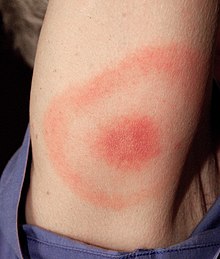
  • The most common sign of infection is an expanding area of redness on the skin, known as erythema migrans [1]
  • Blood test - Screening Lyme serology - positive [2]
  • Lyme carditis is a complication of Lyme disease [3]
  • The Lyme bacterium can infect all parts of the heart, including the conduction system around the atrioventricular node, the outer or inner membranes of the heart, the cardiac muscle [4]
  • impairment of conduction system cause heart block [ 1st degree, 2nd degree and complete heart block]
  • Ceftriaxone started after 24 hours rash is resolved [2]
  • After 48 hours of observation, he underwent a temporary VVI (ventricular pacing and sensing mode) pacemaker insertion [2]
  • On ceftriaxone, the AV block converted to first degree 2 days after starting antibiotics and then completely resolved by day 11, at which point the pacemaker was removed [2]
  • He completed a total 21-day course of treatment with oral doxycycline [2]


References

  1. Lyme disease - Wikipedia
  2. Case of Reversible Complete Heart Block - The American Journal of Medicine, Vol 130, No 8, August 2017
  3. Lyme carditis with complete heart block: management with an external pacemaker, Muhammad Ali Chaudhry. Pubmed
  4. Lyme Disease and the Heart. - Peter J. Krause, Linda K. Bockenstedt. Circulation, 2013


Sunday, September 24, 2017

Brugada syndrome - types

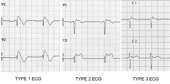
Look at the image, then follow the text.

Look at the right pericordial leads (V1-V3)
Brugada type 1: 
1) Coved type ST elevation
2) J point or ST elevation > 2mm
3) Negative T wave
Brugada type 2: 
1) saddle-back appearance
2) J point elevation > 2mm, ST elevation > 1mm
3) Positive or Bipshasic T wave
Brugada type 3:
1) saddleback or coved appearance
2) ST-segment elevation <1 mm


Tuesday, March 28, 2017

Tilt-table test

  1. is used to indentify cause of SENCOPE
  2. we will identify cause of sencope: vasodepressor or cardioinhibitory
  3. patient lay on table in the supin position
  4. tilting the table maximum: 60-80degrees for 25-40minute
  5. What is result? - Nothing? Lets repeat test with drugs which provoke sencope.
  6. Which drugs we can use? - sublingual nitroglycerin, epinephrine, adenosine, mostly-isoproterenol
  7. Role of isoproterenol: - induces a vasodepressor (decrease in heart rate and blood pressure)
  8. Neurocardyogenic sencopes: Vasodepressor, Carboinhibitory, Mixed
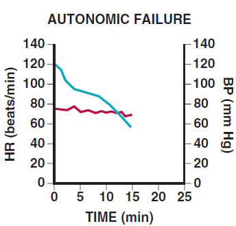
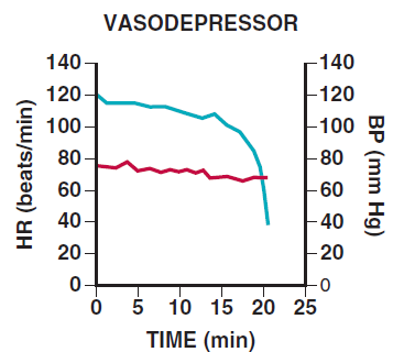
  9. What results can be?: Autonomic failure, Postural Tachycardia, Vasodepresor,
    RESPONSES

    a normal response is an early, slight drop in BP with a compensatory increase in HR mediated by the autonomic nervous system.


    With autonomic dysfunction, a progressive fall in BP is not counteracted by an increase in HR

    In postural tachycardia syndrome, an exaggerated increase in HR is seen


    Vasodepressor is a type of neurocardiogenic sencope. Pure vasodepressor response is a relatively sudden drop in BP without a marked change in HR

    cardioinhibitory is also neurocadiogenic sencope. Cardioinhibitory response shows a sudden decrease in HR without a change in BP

    A mixed response shows decreases in both HR and BP.(also neurocardiogenic sencope)

Sunday, February 26, 2017

Why we review the serum creatinine?

Serum creatinine

1) Liver produce creatinine
2) Creatin kinase ferment phosphorylate creatinine to: phosphocreatine
3) phosphocreatine is high-energy compound so muscle and brain need it mainly
4) at the end of metabolysm kidnet remove creatinin
5) if the function of kidneys funstion slows down then creatinine level will increase

Do not be envious! Share your knowledge with us. Comment more information. Share also..

Right parasternal position

Do you use right parasternal position on echocardiography? If we dont use then why we say that position while talking about echocardiography?

This position is useful to examine the aorta or interatrial septum and is also useful in patients with congenital malposition of the heart, such as dextrocardia. If you have more information do not be envious. Share your knowledge with us.

Friday, February 24, 2017

Congenital Heart Defects

Lets put the list of CHD here, then step by step review them :)
Aortic stenosis
Atrial septal defect (ASD)
Atrioventricular septal defect (AVSD)
Bicuspid aortic valve
Dextrocardia
Double inlet left ventricle (DILV)
Double outlet right ventricle (DORV)
Ebstein's anomaly
Hypoplastic left heart syndrome (HLHS)
Hypoplastic right heart syndrome (HRHS)
Mitral stenosis
Pulmonary atresia
Pulmonary stenosis
Transposition of the great vessels
dextro-Transposition of the great arteries (d-TGA)
levo-Transposition of the great arteries (l-TGA)
Tricuspid atresia
Persistent truncus arteriosus
Ventricular septal defect (VSD)
Wolff-Parkinson-White syndrome (WPW)
Some conditions affect the great vessels or other vessels in close proximity to the heart, but not the heart itself, but are often classified as congenital heart defects.
Coarctation of the aorta (CoA)
Double aortic arch, aberrant subclavian artery, and other malformations of the great arteries
Interrupted aortic arch (IAA)
Patent ductus arteriosus (PDA)
Scimitar syndrome (SS)
Partial anomalous pulmonary venous connection (PAPVC)
Total anomalous pulmonary venous connection (TAPVC)
Some constellations of multiple defects are commonly found together.
tetralogy of Fallot (ToF)
pentalogy of Cantrell
Shone's syndrome/ Shone's complex / Shone's anomaly

Ventricular septal defect types

How many VSD types do you know? Lets count together..
1. Perimembranous VSD
2. Muscular VSD
3. Inlet VSD
4. Infundibular (supracristal) VSD
5. Apical VSD


Watch the video for more information about VSD

Thursday, February 16, 2017

Carcinoid Heart Disease

1) Fixed tricuspid valve
2) Dilated RA and RV
3) Carcinoid tumors are neuroendocrine malignancies
4) Carcinoid tumors secrete large amounts of vasoactive substances: (5-hydroxytryptamine, tachykinins, prostaglandins)
5) Carcinoid SYNDROME occurs when tumor cells metastasize to the liver
6)  Vasoactive substances start to releasing from liver
7) That substances reaching the right heart (RA, RV)
8) deposition of fibrous tissue on endocard


Monday, January 9, 2017

Junctional Tachycardia

Q: What is Junctional Tachycardia?
A: Its a type of SVT
Q: What is the pathology?
A: SA node automaticity decrease and AV node pacemaker exceed SA node.
Q: Can you say the causes?
A: Digoxin toxicity, coronary syndrome, beta-agonists, myocarditis, heart failure
Q: Good, do you know it types?
A: Oh no, i fotgot :(
Q: It has three types
1) Juctional Escape Rhythm - 40-60bpm
2) Juctional Accelerated Rhythm - 60-100bpm
3) Junctional Tachycardia > 100bpm
Q: How you will know Junctional Rhythm on ECG paper?
A: i dont know(


Q: Firstly we should know that whats happening if AV node exceed SA node. Read attentively. If AV node exceed SA node then, impuls will start from AV node so impuls go up to depolarisation of auricularies. In normal cases impuls start from top to down + left to right so P wave is positive in II lead. So in junctional rhythm it will be negative.
A: Let me ask a question. How can we difference Junctional Tachycardia and VT?


Saturday, January 7, 2017

Sinus Node Dysfunction, Sick Sinus Syndrome, Sinus Arrest

Q: What do you say about P waves for this ECG?
A: P waves i think normal.
Q: and PR interval?
A: It seems PR interval also normal between 0.12-0.20sec
Q: and QRS complex?
A: i dont see any patology, its also between 0.06-0.20sec
Q: What about rhythm?
A: Hmm, its irregular
Q: You are right. The pathology in this case is Sinus arrest. Look attentively P wave pause is more than 3 second. Its a Sinus Node Dysfunction. Also you we can say: Sick Sinus Syndrome

Tuesday, December 27, 2016

Marfan syndrome

Q: Tell me about Marfan syndromeA: genetically disorders of connective tissueQ: Which gen?A: FBN1 gen, which has role in producing of fibrillin-1Q: Whats the main complications in cardiovascular system?A: mitral valve prolaps and aortic aneurysmQ: What is Bentall procedure?A: Bentall procedure is composite cardiac surgery operationQ: Which surgery operations involving?A:  Replacement of the aortic valveaortic root and ascending aorta, with re-implantation of the coronary arteriesQ: in which case Bentall surgery is used?A:This operation is used to treat combined aortic valve and ascending aorta disease, including lesions associated with Marfan syndrome.